Subscribe

DOI: 10.4244/EIJV9I10A210

Signet ring-like appearance: specific feature of vulnerable plaques detected by 320-slice multidetector computed tomography

Hironori Hara, MD; Jiro Aoki*, MD, PhD, FACC, FAHA, FESC; Kengo Tanabe, MD, PhD; Masanori Taniwaki, MD; Kazuhiro Hara, MD

A 59-year-old man presented to the emergency department with sustained chest pain. Emergency coronary angiography (CAG) revealed an occluded right coronary artery (RCA) (Figure 1A). After aspiration, a coronary stent was implanted. Histopathological examination showed that the thrombus contained cholesterol crystals and foam cells (Figure 1B and Figure 1C). He was discharged 11 days later without any complications.

A 320-slice multidetector computed tomography (CT) had been accidentally performed five days prior to plaque rupture. He had been suffering from stable angina with ST depression in leads II, III, and aVF, and without elevation of cardiac enzymes. He preferred non-invasive coronary CT to CAG at first. CT images demonstrated moderate luminal narrowing in the RCA (Figure 1D, Figure 1E and Figure 1F) and the lesion had a large calcification, which associated with stable plaque (Figure 1G). Notably, we observed a specific type of cross-sectional image, the so-called signet ring-like appearance (Figure 1H).

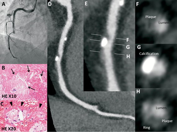
Figure 1. Angiography showed complete occlusion of the middle RCA (A). The thrombus contained cholesterol crystals (arrows, B) and foam cells (arrowheads, C). The images showed the multiplanar reconstruction image of the RCA (D and E). The lumen was moderately narrowed by a moderate CT density plaque (60 HU in the cross-sectional view, F) and the plaque contained a focal large calcification (G). The signet ring-like appearance, defined as a semicircular thin enhancement, was observed along the outer contour of the vessel wall (H).

This appearance has been defined as a semicircular thin enhancement (CT density >130 HU) along the outer contour of the vessel wall in cross-sectional views1. This appearance might indicate deep calcification within lipid-rich plaque containing a necrotic core or intraplaque vasa vasorum, which is one of the specific features of vulnerable plaque.

Conflict of interest statement

The authors have no conflicts of interest to declare.

Volume 9 Number 10
Feb 20, 2014
Volume 9 Number 10
View full issue


Key metrics

Suggested by Cory

10.4244/EIJV16I5A65 Aug 28, 2020
OCT-derived coronary calcified nodules as a predictor of high-risk patients
Akasaka T and Kubo T
free

10.4244/EIJV15I9A138 Oct 4, 2019
Ruptured and healed atherosclerotic plaques: breaking bad?
Amabile N and Veugeois A
free

Editorial

10.4244/EIJ-E-22-00051 Jan 23, 2023
What makes a plaque rupture? A simple answer seems too much to ask for
Achenbach S
free

IMAGE IN CARDIOLOGY

10.4244/EIJV11I4A86 Aug 20, 2015
Calcified nodule: a double paradox on coronary imaging
Alfonso F et al
free

INTERVENTIONAL FLASHLIGHT

10.4244/EIJ-D-17-00087 Jun 2, 2017
Acute myocardial infarction in a young endurance athlete caused by probable plaque erosion
Colleran R et al
free

State-of-the-Art

10.4244/EIJ-D-24-00387 Jul 21, 2025
Advances in coronary imaging of atherosclerotic plaques
Garcia-Garcia H et al
free

State-of-the-Art

10.4244/EIJ-D-25-00296 Dec 1, 2025
Identification and treatment of calcified nodules in percutaneous coronary intervention
Ali Z et al
free

Debate

10.4244/EIJ-E-23-00002 Jun 5, 2023
All calcified nodules are made equal and require the same approach: pros and cons
Guagliumi G et al
free

IMAGE IN CARDIOLOGY

10.4244/EIJV10I12A251 Apr 20, 2015
Very late stent thrombosis caused by neoatherosclerotic plaque rupture
Hougaard M et al
free
Chat with Cory
Hello , I'm Cory and I will do my best to answer your questions about this article. Please remember that this is an experimental feature, and that I'm still learning.
How did the findings from the CT imaging compare to the subsequent angiographic findings?
What are the implications of this case for clinical decision-making and targeted interventions to prevent acute coronary syndromes?
What are the potential limitations or challenges in using CT imaging to assess plaque vulnerability?
What additional diagnostic or prognostic information could have been obtained from this case to further elucidate the mechanisms of plaque rupture and acute coronary syndrome?
X

PCR
Impact factor: 9.5
2024 Journal Citation Reports®
Science Edition (Clarivate Analytics, 2025)
Online ISSN 1969-6213 - Print ISSN 1774-024X
© 2005-2026 Europa Group - All rights reserved